CT Diagnosis of Lupus Enteritis
Article Information
Lokesh Singh MD1, Aruna Singh MD2*
1Department of Radio diagnosis, PGIMER, Chandigarh, India
2Department of Obstetrics and Gynecology, PGIMER, Chandigarh, India
*Corresponding Author: Aruna Singh MD, Department of Obstetrics and Gynecology, PGIMER, Chandigarh, India
Received: 23 October 2020; Accepted: 09 November 2020; Published: 02 December 2020
Citation: Lokesh Singh, Aruna Singh. CT Diagnosis of Lupus Enteritis. Archives of Clinical and Medical Case Reports 4 (2020): 1162-1164.
View / Download Pdf Share at FacebookKeywords
Lupus enteritis; Abdominal pain
Lupus enteritis articles; Abdominal pain articles
Lupus enteritis articles Lupus enteritis Research articles Lupus enteritis review articles Lupus enteritis PubMed articles Lupus enteritis PubMed Central articles Lupus enteritis 2023 articles Lupus enteritis 2024 articles Lupus enteritis Scopus articles Lupus enteritis impact factor journals Lupus enteritis Scopus journals Lupus enteritis PubMed journals Lupus enteritis medical journals Lupus enteritis free journals Lupus enteritis best journals Lupus enteritis top journals Lupus enteritis free medical journals Lupus enteritis famous journals Lupus enteritis Google Scholar indexed journals Abdominal pain articles Abdominal pain Research articles Abdominal pain review articles Abdominal pain PubMed articles Abdominal pain PubMed Central articles Abdominal pain 2023 articles Abdominal pain 2024 articles Abdominal pain Scopus articles Abdominal pain impact factor journals Abdominal pain Scopus journals Abdominal pain PubMed journals Abdominal pain medical journals Abdominal pain free journals Abdominal pain best journals Abdominal pain top journals Abdominal pain free medical journals Abdominal pain famous journals Abdominal pain Google Scholar indexed journals DNA articles DNA Research articles DNA review articles DNA PubMed articles DNA PubMed Central articles DNA 2023 articles DNA 2024 articles DNA Scopus articles DNA impact factor journals DNA Scopus journals DNA PubMed journals DNA medical journals DNA free journals DNA best journals DNA top journals DNA free medical journals DNA famous journals DNA Google Scholar indexed journals Pleural biopsy articles Pleural biopsy Research articles Pleural biopsy review articles Pleural biopsy PubMed articles Pleural biopsy PubMed Central articles Pleural biopsy 2023 articles Pleural biopsy 2024 articles Pleural biopsy Scopus articles Pleural biopsy impact factor journals Pleural biopsy Scopus journals Pleural biopsy PubMed journals Pleural biopsy medical journals Pleural biopsy free journals Pleural biopsy best journals Pleural biopsy top journals Pleural biopsy free medical journals Pleural biopsy famous journals Pleural biopsy Google Scholar indexed journals CECT articles CECT Research articles CECT review articles CECT PubMed articles CECT PubMed Central articles CECT 2023 articles CECT 2024 articles CECT Scopus articles CECT impact factor journals CECT Scopus journals CECT PubMed journals CECT medical journals CECT free journals CECT best journals CECT top journals CECT free medical journals CECT famous journals CECT Google Scholar indexed journals treatment articles treatment Research articles treatment review articles treatment PubMed articles treatment PubMed Central articles treatment 2023 articles treatment 2024 articles treatment Scopus articles treatment impact factor journals treatment Scopus journals treatment PubMed journals treatment medical journals treatment free journals treatment best journals treatment top journals treatment free medical journals treatment famous journals treatment Google Scholar indexed journals CT articles CT Research articles CT review articles CT PubMed articles CT PubMed Central articles CT 2023 articles CT 2024 articles CT Scopus articles CT impact factor journals CT Scopus journals CT PubMed journals CT medical journals CT free journals CT best journals CT top journals CT free medical journals CT famous journals CT Google Scholar indexed journals surgery articles surgery Research articles surgery review articles surgery PubMed articles surgery PubMed Central articles surgery 2023 articles surgery 2024 articles surgery Scopus articles surgery impact factor journals surgery Scopus journals surgery PubMed journals surgery medical journals surgery free journals surgery best journals surgery top journals surgery free medical journals surgery famous journals surgery Google Scholar indexed journals Cardioverter articles Cardioverter Research articles Cardioverter review articles Cardioverter PubMed articles Cardioverter PubMed Central articles Cardioverter 2023 articles Cardioverter 2024 articles Cardioverter Scopus articles Cardioverter impact factor journals Cardioverter Scopus journals Cardioverter PubMed journals Cardioverter medical journals Cardioverter free journals Cardioverter best journals Cardioverter top journals Cardioverter free medical journals Cardioverter famous journals Cardioverter Google Scholar indexed journals enterectomy articles enterectomy Research articles enterectomy review articles enterectomy PubMed articles enterectomy PubMed Central articles enterectomy 2023 articles enterectomy 2024 articles enterectomy Scopus articles enterectomy impact factor journals enterectomy Scopus journals enterectomy PubMed journals enterectomy medical journals enterectomy free journals enterectomy best journals enterectomy top journals enterectomy free medical journals enterectomy famous journals enterectomy Google Scholar indexed journals
Article Details
1. Case Report
A 25 year old female patient was a diagnosed case of systemic lupus erythematosus with haematological, musculoskeletal, oral and dermatological manifestations. Patient was stable and remained asymptomatic for last 5 years. For last 2 months as the steroids were tapered and patient missed few doses, she developed fever, joint pains, oral ulcers, odynophagia and shortness of breath. Patient also complained of abdominal pain which was acute in onset, moderate to severe in intensity, continuous, diffuse involving whole of abdomen and relieved partially on analgesic. There were 2-3 episodes of vomiting followed by loose stools. No h/o hematemesis or melena was seen.
On examination, pulse 132 b/min, BP 140/90 mm Hg, SpO2-97% on room air, RR- 28/min. patient was conscious and alert. No pallor, icterus, cyanosis, clubbing, LAP/edema seen. On local examination-mild tenderness present over right hypochondrium. Bowel sounds were sluggish. No organomegaly/ free fluid. Initially laboratory workup showed anaemia. LFT, RFT, coagulation profile were within normal limits. Serological examination showed elevated ANA, Anti ds DNA, raised hsDNA and raised pro-BNP.
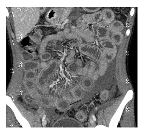
For evaluation of abdominal pain, plain X-ray of abdomen was done which was normal. Ultrasound scan of abdomen showed mild amount of free fluid in abdomen and pelvis. For further evaluation of patient, CT angiography of abdominal vessels followed by CECT of abdomen was done which showed small bowel wall thickening and enhancement giving characteristic ‘target sign’ (formed by enhancing innermost mucosa, middle non enhancing submucosa and outermost enhancing muscularis propria ± serosa) (Figure 1). There was adjacent mesenteric fat stranding with prominent mesenteric vasculature giving comb sign (Figure 1). Mild ascites was also seen. Abdominal aorta and major branches were normal in contrast opacification. Based on these characteristic features, diagnosis of lupus enteritis was kept. Patient was started on parentral high dose steroid therapy following which marked improvement was observed in symptoms.

Figure 1: Coronal Post contrast CT image showing dilated and thickened small bowel loops (target sign, black arrow) with mesenteric vessel engorgement (comb sign, white arrow).
Lupus enteritis is a relatively uncommon manifestation of SLE and frequently presents as abdominal pain, diarrhoea and vomiting [1]. It most commonly involves the jejunum and ileum. Principal imaging findings seen on abdominal CT are bowel wall thickening > 3 mm also called as target sign, engorgement of mesenteric vessels also known as comb sign and increased attenuation of mesenteric fat [2]. Treatment for lupus enteritis is high-dose intravenous steroid therapy and complete bowel rest. In few cases immunosuppressants such as azathioprine or cyclophosphamide may be beneficial [3].
Conflicts of Interest
Nil
Funding
Nil
References
- Zizic TM, Shulman LE, Stevens MB. Colonic perforations in systemic lupus erythematosus. Medicine (Baltimore) 54 (1975): 411-426.
- Lee CK, Ahn MS, Lee EY, et al. Acute abdominal pain in systemic lupus erythematosus: focus on lupus enteritis (gastrointestinal vasculitis). Ann Rheum Dis 61 (2002): 547-550.
- Sultan SM, Ioannou Y, Isenberg DA. A review of gastrointestinal manifestations of systemic lupus erythematosus. Rheumatology (Oxford) 38 (1999): 917-932.


 Impact Factor: * 3.1
 CiteScore: 2.9
  Acceptance Rate: 11.01%
  Time to first decision: 10.4 days
  Time from article received to acceptance: 2-3 weeks
