Gestational Gigantomastia
Article Information
Aruna Singh MD1, Lokesh Singh MD2, Ashima Arora MD1*
1Department of Obstetrics and Gynecology, PGIMER, Chandigarh, India
2Department of Radio diagnosis, PGIMER, Chandigarh, India
*Corresponding Author: Ashima Arora MD, Department of Obstetrics & Gynaecology, PGIMER, Chandigarh, India
Received: 23 October 2020; Accepted: 09 November 2020; Published: 07 December 2020
Citation: Aruna Singh, Lokesh Singh, Ashima Arora. Gestational Gigantomastia. Archives of Clinical and Medical Case Reports 4 (2020): 1208-1210.
View / Download Pdf Share at FacebookKeywords
Gestational Gigantomastia; Enlarged breasts
Gestational Gigantomastia articles; Enlarged breasts articles
Gestational Gigantomastia articles Gestational Gigantomastia Research articles Gestational Gigantomastia review articles Gestational Gigantomastia PubMed articles Gestational Gigantomastia PubMed Central articles Gestational Gigantomastia 2023 articles Gestational Gigantomastia 2024 articles Gestational Gigantomastia Scopus articles Gestational Gigantomastia impact factor journals Gestational Gigantomastia Scopus journals Gestational Gigantomastia PubMed journals Gestational Gigantomastia medical journals Gestational Gigantomastia free journals Gestational Gigantomastia best journals Gestational Gigantomastia top journals Gestational Gigantomastia free medical journals Gestational Gigantomastia famous journals Gestational Gigantomastia Google Scholar indexed journals Gigantomastia articles Gigantomastia Research articles Gigantomastia review articles Gigantomastia PubMed articles Gigantomastia PubMed Central articles Gigantomastia 2023 articles Gigantomastia 2024 articles Gigantomastia Scopus articles Gigantomastia impact factor journals Gigantomastia Scopus journals Gigantomastia PubMed journals Gigantomastia medical journals Gigantomastia free journals Gigantomastia best journals Gigantomastia top journals Gigantomastia free medical journals Gigantomastia famous journals Gigantomastia Google Scholar indexed journals Enlarged breasts articles Enlarged breasts Research articles Enlarged breasts review articles Enlarged breasts PubMed articles Enlarged breasts PubMed Central articles Enlarged breasts 2023 articles Enlarged breasts 2024 articles Enlarged breasts Scopus articles Enlarged breasts impact factor journals Enlarged breasts Scopus journals Enlarged breasts PubMed journals Enlarged breasts medical journals Enlarged breasts free journals Enlarged breasts best journals Enlarged breasts top journals Enlarged breasts free medical journals Enlarged breasts famous journals Enlarged breasts Google Scholar indexed journals oligohydraminos articles oligohydraminos Research articles oligohydraminos review articles oligohydraminos PubMed articles oligohydraminos PubMed Central articles oligohydraminos 2023 articles oligohydraminos 2024 articles oligohydraminos Scopus articles oligohydraminos impact factor journals oligohydraminos Scopus journals oligohydraminos PubMed journals oligohydraminos medical journals oligohydraminos free journals oligohydraminos best journals oligohydraminos top journals oligohydraminos free medical journals oligohydraminos famous journals oligohydraminos Google Scholar indexed journals primigravida articles primigravida Research articles primigravida review articles primigravida PubMed articles primigravida PubMed Central articles primigravida 2023 articles primigravida 2024 articles primigravida Scopus articles primigravida impact factor journals primigravida Scopus journals primigravida PubMed journals primigravida medical journals primigravida free journals primigravida best journals primigravida top journals primigravida free medical journals primigravida famous journals primigravida Google Scholar indexed journals treatment articles treatment Research articles treatment review articles treatment PubMed articles treatment PubMed Central articles treatment 2023 articles treatment 2024 articles treatment Scopus articles treatment impact factor journals treatment Scopus journals treatment PubMed journals treatment medical journals treatment free journals treatment best journals treatment top journals treatment free medical journals treatment famous journals treatment Google Scholar indexed journals CT articles CT Research articles CT review articles CT PubMed articles CT PubMed Central articles CT 2023 articles CT 2024 articles CT Scopus articles CT impact factor journals CT Scopus journals CT PubMed journals CT medical journals CT free journals CT best journals CT top journals CT free medical journals CT famous journals CT Google Scholar indexed journals surgery articles surgery Research articles surgery review articles surgery PubMed articles surgery PubMed Central articles surgery 2023 articles surgery 2024 articles surgery Scopus articles surgery impact factor journals surgery Scopus journals surgery PubMed journals surgery medical journals surgery free journals surgery best journals surgery top journals surgery free medical journals surgery famous journals surgery Google Scholar indexed journals SARS-COV-2 articles SARS-COV-2 Research articles SARS-COV-2 review articles SARS-COV-2 PubMed articles SARS-COV-2 PubMed Central articles SARS-COV-2 2023 articles SARS-COV-2 2024 articles SARS-COV-2 Scopus articles SARS-COV-2 impact factor journals SARS-COV-2 Scopus journals SARS-COV-2 PubMed journals SARS-COV-2 medical journals SARS-COV-2 free journals SARS-COV-2 best journals SARS-COV-2 top journals SARS-COV-2 free medical journals SARS-COV-2 famous journals SARS-COV-2 Google Scholar indexed journals enterectomy articles enterectomy Research articles enterectomy review articles enterectomy PubMed articles enterectomy PubMed Central articles enterectomy 2023 articles enterectomy 2024 articles enterectomy Scopus articles enterectomy impact factor journals enterectomy Scopus journals enterectomy PubMed journals enterectomy medical journals enterectomy free journals enterectomy best journals enterectomy top journals enterectomy free medical journals enterectomy famous journals enterectomy Google Scholar indexed journals
Article Details
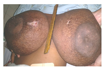
A 28-year-old primigravida with oligohydraminos and IUGR presented for her first prenatal visit at 35 weeks of gestation with complains of severe back pain, marked breast enlargement and tenderness. The patient reported that breast enlargement started at 5 months of gestation and was the first time she had experienced excessive breast enlargement. No significant past medical history was present. She reported that her breast size prior to pregnancy was around 32B and estimates her breasts are now 10 times bigger during this pregnancy. Physical exam revealed bilateral enlarged breasts with presence of an ulcer of 3-4 cm over right upper inner quadrant (Figure 1). Serum prolactin levels were normal. Other laboratory investigations were also unremarkable. Ultrasound of breast revealed skin thickening and dilated ducts and vascular structures. A biopsy was performed from the right breast which suggested atypical lobular hyperplasia. Patient refused for breast reduction surgery. Planned induction of labour was done at 37 weeks, however it was converted to caesarean section due to fetal distress. A live born boy with weight of 2.2 kg was delivered. No complications were seen. Patient was discharged and kept on follow up with reduction of up to sixty percent in size of breasts after 6 weeks post partum.

Figure 1: Clinical image of patient showing massively enlarged breasts with ulceration.
Gestational gigantomastia is a rare and debilitating condition which is characterized by massive enlargement of the breasts with associated tissue necrosis or haemorrhage and psychosocial implications [1, 2]. GG is an uncommon disease and the exact pathophysiology is not clear. The risk factors reported are Caucasian women, multiparity, occurrence in prior pregnancy and association with autoimmune diseases [2, 3]. The majority of cases are bilateral and seen in early pregnancy. The condition is benign and may mimic malignancy due to rapid enlargement and skin edema. Sometimes a biopsy should be done if high clinical suspicion of malignancy is seen [3].
Treatment is still controversial and modalities include medical management with bromocriptine and surgical breast reduction. In our case, the patient opted for watchful observation without treatment and was kept on follow up. There was significant reduction in breast size at a 6 month follow up.
Conflicts of Interest
Nil
Funding
Nil
References
- Swelstad MR, Swelstad BB, Rao VK, et al. Management of gestational gigantomastia. Plast Reconstr Surg 118 (2006): 840-848.
- Dem A, Wone H, Faye ME, et al. Bilateral gestational macromastia: case report. J Gynecol Obstet Biol Reprod (Paris) 38 (2009): 254-257.
- Mangla M, Singla D. Gestational Gigantomastia: A Systematic Review of Case Reports. J Midlife Health 8 (2017): 40-44.


 Impact Factor: * 3.1
 CiteScore: 2.9
  Acceptance Rate: 11.01%
  Time to first decision: 10.4 days
  Time from article received to acceptance: 2-3 weeks
