Prevalence and Distribution of Odontogenic Cysts and Tumors in North Indian Population: A Database Study with Systematic Review
Article Information
Jaya Singh1, Shruti Singh1, Shaleen Chandra2, Fahad M. Samadi3*
1Senior Resident, Department of Oral Pathology and Microbiology, Faculty of Dental Sciences, KGMU, Lucknow, India
2Professor and Head, Department of Oral Pathology and Microbiology, Faculty of Dental Sciences, KGMU, Lucknow, India
3Associate Professor, Department of Oral Pathology and Microbiology, Faculty of Dental Sciences, KGMU, Lucknow, India
*Corresponding Author: Fahad M. Samadi, Associate Professor, Department of Oral Pathology and Microbiology, King George’s Medical University, Lucknow, UP, India
Received: 27 April 2020; Accepted: 08 May 2020; Published: 13 May 2020
Citation: Jaya Singh, Shruti Singh, Shaleen Chandra, Fahad M. Samadi. Prevalence and Distribution of Odontogenic Cysts and Tumors in North Indian Population: A Database Study with Systematic Review. International Journal of Applied Biology and Pharmaceutical Technology 11 (2020): 46-59.
View / Download Pdf Share at FacebookAbstract
Abstract
Aim and Objective: The aim of this study was to determine the prevalence of odontogenic cysts and tumors over a time period of 5 years.
Materials and Methods: Data for this analysis was attained from the reports of patients diagnosed with odontogenic cysts and tumors. Their prevalence and distribution according to age, gender, site and histopathologic types were noted and statistically analyzed.
Results: Among 893 patients visiting the department between years 2014-18, 33 patients presented with odontogenic cysts and 56 patients presented with odontogenic tumors. The prevalent age group was seen ranging from 19 to 40 years for both cysts and tumors. While the cyst was more commonly seen in males, tumors were seen in females. Overall prevalence of cyst was recorded as 3.6% and tumors as 6.2%.
Conclusion: The current study provides the relative distribution of different odontogenic cysts and tumors reported at our institution. Variability in the data with previous studies can be attributed to the diverse demographic factors. Thus, these findings can help us in better understanding of such lesions and ameliorate the diagnosis of odontogenic cysts and tumors.
Keywords
Odontogenic cysts; Odontogenic tumors; Odontogenic keratocyst; Ameloblastoma
Article Details
Introduction
Odontogenic cysts and tumors comprise an unusually sundry group of lesions. All these lesions originate by an aberration in the normal pattern of odontogenesis and thereby reflecting the complex development of oro-dental structures. Few lesions are not totally neoplastic but resultant of any disturbance/alteration in the normal development of tooth. Lesions such as cysts are also tumors only in the broadest sense of the word and do not represent true neoplasms [1].
Cysts of the jaws can be Odontogenic and Non-odontogenic. The odontogenic cysts are derived from epithelium associated with the development of the dental apparatus. The type of epithelium can vary with most lesions having stratified squamous but some developmental or fissural cysts in the maxilla may have respiratory epithelium. Several types of odontogenic cysts may occur, dependent chiefly upon the stage of odontogenesis during which they originate [1].
Odontogenic tumors are derived from ectomesenchymal and/or epithelial tissues that constitute the tooth-forming apparatus [2]. The odontogenic tumors represent inductive interactions between odontogenic ectomesenchyme and epithelium like normal odontogenesis [3, 4]. Therefore odontogenic tumors are found within the jaw bones (central types) or in the mucosal tissue overlying tooth-bearing areas (peripheral types). Odontogenic tumors are divided intotwo primary categories; benign and malignant, the etiology of which is unknown.
Most of the benign odontogenic cysts and tumors arise de novo, whereas the malignant odontogenic cysts and tumors may arise denovo but more often arise from their benign precursor. The classification of odontogenic cysts and tumors isessentially based on interactions between odontogenic ectomesenchyme and epithelium. This dynamic classification is constantly renewed with the addition of new entities, and the removal of some older entities. The last update of these tumors was published in early 2017 [5].
Despite the vast number of cases, studies on odontogenic cysts and tumors are relatively less in the literature and information regarding the demographic profile of these lesions in different populations is scarce. However, the present study was designed to know the relative frequency of odontogenic cysts and tumors and to relate with their clinico-pathological characteristics.
Materials and Methods
All cases of odontogenic cysts and tumors diagnosed histopathologically between 2014 and 2018 were reviewed from the archives of the Department of Oral and Maxillofacial Pathology. The following variables were recorded: gender, age and location. To classify the location of the cysts and tumors, each jaw was divided into an anterior and a posterior zone. The anterior zone included the incisors, canines and premolars, and the posterior zone consisted of the molars and ramus/tuberosity. Otherwise, it was placed in the zone according to radiographic evaluation. The data was analyzed, and graphs were formulated respectively.
Results
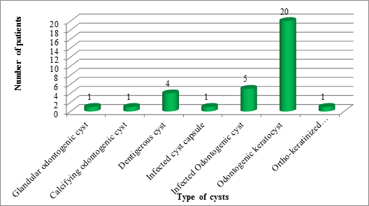
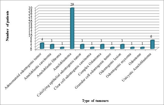
Among 893 patients visiting the department between years 2014-18, 33 patients presented with odontogenic cysts and 56 patients presented with odontogenic tumours. Overall prevalence of cyst was recorded as 3.6% and tumors as 6.2%. In the year 2014, 7 patients presented with cyst and 13 presented with tumours. In the year 2015, 5 patients presented with cyst and 10 presented with tumours. In the year 2016, 4 patients presented with cyst and 10 presented with tumours. In the year 2017, 8 patients presented with cyst and 14 presented with tumours. While in the year 2018, equal number of 9 patients presented with cyst and tumours. Figure 1&2 shows the overall prevalence of cysts and tumors respectively.
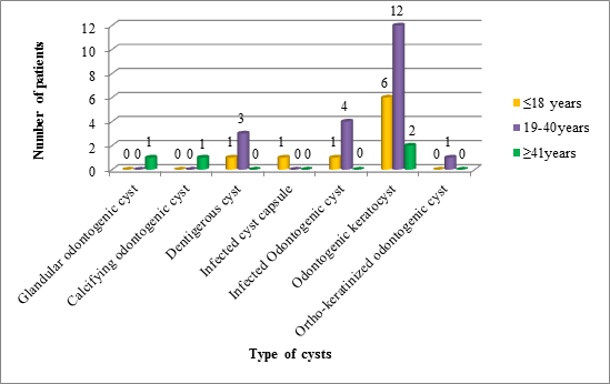
Figure 3 illustrates the comparison of the types of cyst according to age, 12 patients aged 19-40 years, 6 patients in age group of ≤18 years and 2 patients in aged ≥41years presented with Odontogenic keratocyst. However, there was no statistically significant difference among different age groups.
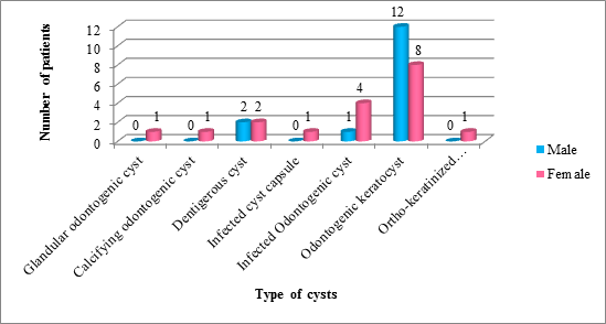
Figure 4 illustrates the comparison of the types of cyst according to gender, 12 male patients and 8 female patients presented with Odontogenic keratocyst. Also, 4 female patients presented with Infected Odontogenic cyst. There was no statistically significant difference among both genders.
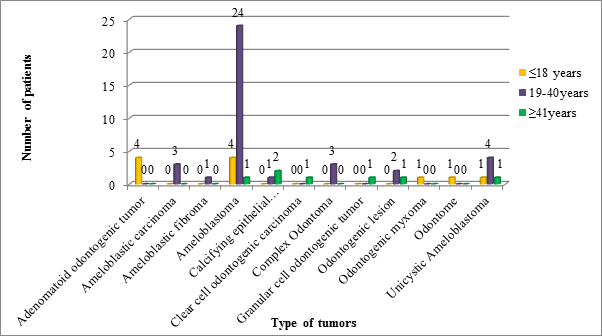
Figure 5 illustrates the comparison of the types of tumours according to age, 24 patients aged 19-40 years, 4 patients in age group of ≤18 years and 1 patient in aged ≥41years presented with Ameloblastoma [4]. patients in age group of ≤18 years presented with Adenomatoid odontogenic tumor and 4 patients in age group of 19-40 years presented with Unicystic Ameloblastoma. There was a statistically significant difference found among different age groups.
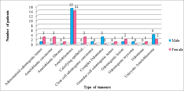
Figure 6 illustrates the comparison of the types of tumors according to gender, 15 male patients and 14 female patients presented with Ameloblastoma. 4 male patients and 2 female patients presented with Unicystic Ameloblastoma. 3 female patients presented with each Adenomatoid odontogenic tumor and Ameloblastic carcinoma. There was no statistically significant difference among both genders.
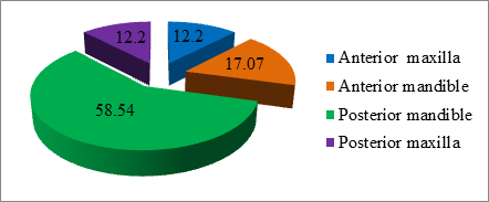
Figure 7 illustrates the presentation of the lesion according to the location. Majority of the patients presented with lesion in the posterior mandible (58.54%) followed by anterior mandible (17.07%).
Table: Distribution of odontogenic cysts and tumors according to prevalence, gender, age and location
Discussion
Odontogenic lesions constitute a group of heterogenous lesions that range from hamartomatous or non-neoplastic tissue proliferations to malignant neoplasm with metastatic capabilities. These lesions are derived from epithelial, ectomesenchymal and/or mesenchymal elements that are, or have been, part of tooth forming apparatus [6]. The diagnosis of odontogenic cysts and tumors should be based on careful examination of clinical, radiographical, and histopathological features. Most of the information regarding the prevalence of odontogenic cysts and tumors comes from diagnostic services and despite some sampling bias; these services represent a reliable source of information regarding the relative frequency and clinical-pathologic features of odontogenic cysts and tumors [7, 8].
Out of the total 893 patients, 3.6% of odontogenic cysts and 6.2% of odontogenic tumors were seen. This was in concordance with studies done by Deepthi et al [9] and Pandiar et al [10]. In the current study, odontogenic cysts were more common in females which is in concordance with a brazilian study [11] and in contrast with Ramachandra et al [12] and Kambalinath et al [13] and odontogenic tumors were also prevalent in females which is in agreement with Ramachandra et al [12] and Rafael et al [14]. Unlike our study, other literatures show a male preponderance [9,10,15,16,17,18]. According to a study done by Varkhede et al [19], males and females are seen to be affected equally. This disparity can be attributed to demographic variation. Mandible is the most frequently affected site, which is contrary with the studies of Ochsenius et al. [7] and de Souza et al [11].
Odontogenic keratocyst (OKC) is the most common odontogenic cyst in our study with a mean prevalence of 60.61%. This is consistent with Rubini et al (66.8%) [16], Pandiar et al (35.9%) [10], Rafael et al (31.6%) [14] and Avelar et al (30%) [20]. Earlier, according to the WHO 2005 classification, OKC was termed as Keratocystic odontogenic tumor (KCOT) owing to its aggressive behavior. Later in the recent classification, WHO in 2017 re- termed KCOT as OKC [5]. In the present study, we have considered OKC as a cystic lesion. OKC was more commonly seen in the wide age range of 19-40 years that is in concordance with various other studies [9,10,12,15,17,20]. A definite male predilectionwas seen in this study similar to the findings of Ramachandra et al [12], Kambalimath et al [13], Rubini et al [16] and Pandiar et al [10] and differed with Mehengi et al [15], Deepthi et al [9], Nalabou et al [17], Rafael et al [14] and Avelar et al. The most common site was the posterior mandible as seen in Kambalimath et al [13], Deepthi et al [9], Nalabou et al [17], Rafael et al [14] and Avelar et al [20]. A variant of OKC i.e. Orthokeratinized Odontogenic Cyst (OOC) showed a mean prevalence of 3.03%. Similar to OKC, OOC was also common in 2nd to 4th decade but showed a female predilection. None of the previous studies have mentioned OOC as a separate entity.
Dentigerous cyst (DC) ranks second amongst all the odontogenic cysts in this study showing mean prevalence of 12.12%. Similar finding was seen by Deepthi et al (17.2%) [9]. However, Kambalimath et al [13] and Ramachandra et al [12] found DC to be the most common odontogenic cyst in their study. The mean prevalence of DC was seen in 2nd to 4th decade, one case was seen in the 1st decade. Our finding was consistent with the study done by Ramachandra et al [12] whereas other studies showed more prevalence in the 1st and 2nd decade [9,13]. Equal predilection of male and female is seen in our literature. This is in contrast with various previous literatures where a male preponderance was seen [9, 12, 13].
Other cysts like Glandular odontogenic cyst (GOC) and Calcifying odontogenic cyst (COC) showed equal prevalence i.e. 3.03%. GOC and COC were more common in 4th to 5th decade, with a definite female predilection. Similar findings were seen by Kambalimath et al [13] but showed a male predilection.
Ameloblastoma is found to be the most prevalent odontogenic tumor with the mean prevalence of 51.79%. Many of the studies obtained the same result [9,12,15,17,18,19]. The peak prevalence was seen in 2nd to 4th decade. This is consistent with various other literatures [9,10,12,14,15,17,18,20]. Male and female were affected equally (1.07:1). Contradictory findings were seen in various studies with few studies showing male predominance [10,12,13,16,20] while others showed a female predilection [9,14,15]. The most frequent site reported was posterior mandible which in consistent with many studies [9,10,14,17,18,19,20]. In our series, we have taken a variant unicystic ameloblastoma as a separate entity. It showed a prevalence of 10.71% seen more in 2nd to 4th decade. Males were affected more than females and posterior mandible was the predominant site.
Adenomatoid odontogenic tumor (AOT) is the second most common odontogenic tumor found in our study with a prevalence rate of 7.14%. This was in consensus with a study done by Ramachandra et al [12], which showed a prevalence of 7.8%. Only one study showed a slightly higher prevalence of the lesion [19]. Various other literatures exhibit a lower prevalence rate [9,10,14,16,17,18]. All the cases were seen at a younger age in this study (<18 years of age). Majority of the literature showed a similar finding [9,10,14,17,18,19,20]. However, Ramachandra et al found a bimodal age distribution [12]. On the other hand, Rubini et al found AOT more common in the 3rd decade [16]. A female preponderance was found which was consistent with [10,18,14,20]. Equal gender distribution was seen in many studies [9,12,16,19] except for Nalabou et al [17] which showed a higher prevalence in males.
Odontoma showed a prevalence rate of 5.36%, which was contradictory to various studies where odontoma showed a higher prevalence rate [9, 10, 12, 14, 15, 18, 19]. Over- or under-reporting has a direct influence on this phenomenon. Most odontomas are discovered on routine radiographs and do not produce clinical symptoms [21]. This may be responsible for the low incidence of odontomas observed in the Indian population, because most patients in our environment do not seek medical consultation unless there are symptoms suggesting an obvious pathology [19]. Odontoma was more commonly seen in 2nd to 4th decade with a definite male predilection.
Calcifying Epithelial Odontogenic Tumor, similar to odontoma, showed a prevalence of 5.36%. It was more commonly seen in 4th to 5th decade affecting the female population more than its counterpart. Ameloblastic Carcinoma also showed a prevalence of 5.36% in our study affecting the middle-aged group (19-40 years of age). All the cases were seen in females. Other studies have shown 3.74% [18], 1.1% [9] and 0.3% [13,14] prevalence rates. All these studies showed the mean age prevalence in 3rd to 6th decade. Majority of the literature showed a male predominance [9,16,18] except Rafael et al [14] which was similar to our finding.
Conclusion
Due to low frequency of odontogenic lesions, only few literatures demonstrate the epidemiological data of odontogenic cysts and tumors. The current study provides the relative distribution of different odontogenic cysts and tumors reported at our institution. Variability in the data with previous studies can be attributed to the diverse demographic factors. Thus, these findings can help us in better understanding of such lesions and ameliorate the diagnosis of odontogenic cysts and tumors. Also, the comparative existence of these cysts and tumors can be scrutinized at a worldwide level to comprehend their prevalence and distribution.
Authors’ Contributions
Dr. Fahad M Samadi envisioned the idea of the current research, Dr. Jaya Singh developed the matter, and prepared the manuscript. Dr. Shruti Singh and Professor Shaleen Chandra reviewed the manuscript. All the authors discussed the conclusion and contributions to the final manuscript.
Conflicts of Interest
All authors have read the journal’s policy on disclosure of potential conflicts of interest and have none to declare.
Source of Funding
The article was self-funded.
References
- Shafer WG, Hine MK, Levy BM, et al. Shafer’s textbook of oral pathology. Diseases of the Skin. Rajendran R, editor 5 (2006): 1103-1107.
- Wright JM, Tekkesin MS. Odontogenic tumors: where are we in 2017?. Journal of Istanbul University Faculty of Dentistry 51 (2017) : S10.
- Nanci A, Ten Cate RA. Embryology of the head, face, and oral cavity. Ten Cate’s Oral Histology: Development, Structure, and Function (2008): 32-56.
- Bilodeau EA, Collins BM. Odontogenic cysts and neoplasms. Surgical pathology clinics 10 (2017): 177-222.
- El-Naggar AK, Chan JK, Grandis JR, et al. WHO classification of Head and Neck Tumours (chapter 8). Odontogenic and maxilofacial bone tumours. 4th ed., IARC: Lyon (2017): 205-260.
- Reichart PA, Philipsen HP. Odontogenic tumors and allied lesions. Quintessence Pub (2004).
- Ochsenius G, Escobar E, Godoy L, et al. Odontogenic cysts: analysis of 2.944 cases in Chile. Medicina Oral, Patología Oral y Cirugía Bucal (Internet) 12 (2007): 85-91.
- Mosqueda-Taylor A, Irigoyen-Camacho ME, Diaz-Franco MA, et al. Odontogenic cysts. Analysis of 856 cases. Medicina oral: organo oficial de la Sociedad Espanola de Medicina Oral y de la Academia Iberoamericana de Patologia y Medicina Bucal 7 (2002): 89-96.
- Deepthi PV, Beena VT, Padmakumar SK, et al. A study of 1177 odontogenic lesions in a South Kerala population. Journal of Oral and Maxillofacial Pathology: JOMFP 20 (2016): 202.
- Pandiar D, Shameena PM, Sudha S, et al. Odontogenic Tumors: A 13-year Retrospective Study of 395 Cases in a South Indian Teaching Institute of Kerala. Oral & Maxillofacial Pathology Journal 6 (2015).
- de Souza LB, Gordon-Nunez MA, Nonaka CF, et al. Odontogenic cysts: demographic profile in a Brazilian population over a 38-year period. Medicina Oral, Patologia Oral y Cirugia Bucal 15 (2010): e583-90.
- Ramachandra S, Shekar PC, Prasad S, et al. Prevalence of odontogenic cysts and tumors: A retrospective clinico-pathological study of 204 cases. SRM Journal of Research in Dental Sciences 5 (2014): 170.
- Kambalimath DH, Kambalimath HV, Agrawal SM, et al. Prevalence and distribution of odontogenic cyst in Indian population: a 10 year retrospective study. Journal of Maxillofacial and Oral Surgery 13 (2014): 10-5.
- Lima-Verde-Osterne R, Turatti E, Cordeiro-Teixeira R, et al. The relative frequency of odontogenic tumors: A study of 376 cases in a Brazilian population. Medicina Oral, Patologia Oral y Cirugia Bucal 22 (2017): e193.
- Mehngi R, Rajendra K, Bhagwat P, et al. Clinical and Histopathological Analysis of Odontogenic Tumors in Institution-A 10 Years Retrospective Study. J Contemp Dent Pract 19 (2018): 1288-1292.
- Rubini C, Mascitti M, Santarelli A, et al. Odontogenic tumors: a retrospective clinicopathological study from two Italian centers. Pathologica 109 (2017): 35-46.
- Nalabolu GR, Mohiddin A, Hiremath SK, et al. Epidemiological study of odontogenic tumours: An institutional experience. Journal of Infection and Public Health 10 (2017): 324-330.
- Taghavi N, Rajabi M, Mehrdad L, et al. A 10-year retrospective study on odontogenic tumors in Iran. Indian Journal of Dental Research 24 (2013): 220.
- Varkhede A, Tupkari JV, Mandale MS, et al. Odontogenic tumors: a review of 60 cases (2010).
- Avelar RL, Antunes AA, de Santana Santos T, et al. Odontogenic tumors: clinical and pathology study of 238 cases. Brazilian Journal of Otorhinolaryngology 74 (2008): 668-673.
- Sriram G, Shetty RP. Odontogenic tumors: a study of 250 cases in an Indian teaching hospital. Oral Surgery, Oral Medicine, Oral Pathology, Oral Radiology, and Endodontology 105 (2008): e14-21.
- Selvamani M, Donoghue M, Basandi PS. Analysis of 153 cases of odontogenic cysts in a South Indian sample population: a retrospective study over a decade. Brazilian Oral Research 26 (2012): 330-334.
- Nadaf A, Farooq S, Khuroo M. Restrospective clinic-pathological study of 106 odontogenic cyst among Kashmiri population. IJCMSR 3 (2018): 53-56.


 Impact Factor: * 3.0
 CiteScore: 2.9
  Acceptance Rate: 11.01%
  Time to first decision: 10.4 days
  Time from article received to acceptance: 2-3 weeks